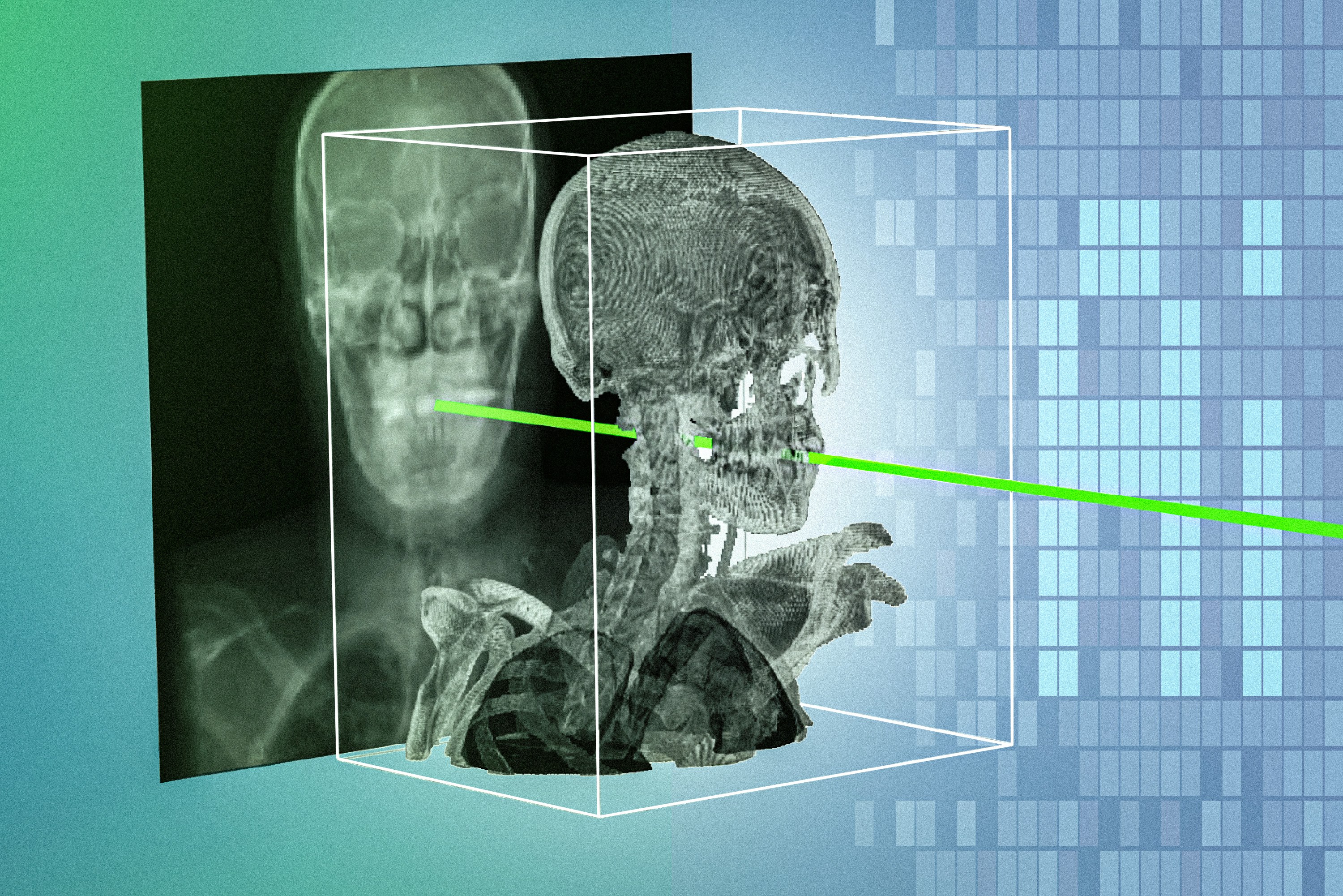
Clinicians performing minimally invasive procedures rely on real-time X-rays to steer surgical instruments, yet flat projection images make it notoriously difficult to determine the precise location and orientation of tools inside the body. While aligning intraoperative 2D images with preoperative 3D volumes like CT or MRI scans solves this localization problem, manual registration remains painfully burdensome. Previous general-purpose deep learning models routinely choked on diverse patient anatomies, blocking their adoption in clinical practice. To bypass this bottleneck, researchers at MIT have developed a technique that rapidly and accurately matches intraoperative X-rays with preoperative 3D scans.
Named xvr (X-ray volume registration), the system takes a single patient's preoperative 3D scan and simulates thousands of synthetic X-rays from multiple angles. It generates synthetic images each second using a physics-based simulation of the X-ray process. While training a registration model from scratch for an individual case would typically take hours, this architecture adapts to each patient in about five minutes.
Sub-Millimeter Precision
During surgery, the tailored model automatically matches the patient's X-rays with the 3D scans in a matter of seconds, achieving sub-millimeter precision. The project was led by Vivek Gopalakrishnan, a postdoc in the MIT Computer Science and Artificial Intelligence Laboratory (CSAIL), along with Polina Golland, professor at MIT, and Neel Dey, investigator at Harvard Medical School and Massachusetts General Hospital. To build and validate the system, the researchers collected whole-body 3D medical scans from more than 1,000 patients.
"We tailor this one specific model for this one specific patient, and it doesn't matter if it works on other people because there will be different models for those people," as Vivek Gopalakrishnan explains.
This shift toward dedicated per-patient adaptation successfully bypasses the generalization failures of earlier models, ensuring robust performance regardless of individual anatomical variation.
Economic and Operational Impact
For healthcare providers and medical device businesses, fast, patient-specific 2D/3D registration demonstrates how targeted simulation can directly cut operating room overhead. By compressing the model adaptation phase to five minutes and alignment time to seconds, the method slashes procedure times, curtails complication risks during catheter and endoscope navigation, and ultimately optimizes operational margins in time-sensitive clinical environments. The primary practical constraint remains the rigid requirement of a prior 3D scan, leaving open questions on how such workflows will integrate into emergency clinical pipelines where preoperative volumetric imaging is absent.