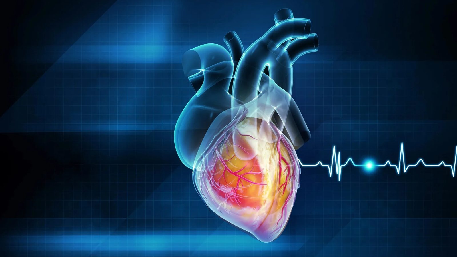
Can clinicians identify sudden cardiac death risk more accurately without deploying complex specialized equipment? Artificial intelligence software trained on routine electrocardiograms can surface severe cardiac risks far more accurately than standard tests, relying entirely on existing clinical infrastructure. By analyzing routine diagnostic traces that medical centers already run every day, the algorithmic screening flags patients at high risk of sudden death who standard evaluations systematically miss.
Rethinking Conventional Metrics
Every year, sudden cardiac arrest kills more than 300,000 people in the United States. The medical emergency occurs when the heart's electrical coordination fails instantly, stopping blood flow.
Roughly 90% of individuals who suffer this event outside a hospital die within minutes. Traditional clinical guidelines attempt to protect vulnerable individuals by evaluating how much blood the heart pumps during each contraction. This metric serves as the primary filter for deciding whether to install an implanted defibrillator. Yet standard clinical metrics isolate a cohort with an annual sudden cardiac death rate of only 4.6%.
Researchers at the University of California - Berkeley set out to solve this diagnostic bottleneck. Published in Nature, their findings reveal a previously overlooked warning signal embedded within standard heart scans. The team trained an artificial intelligence system on more than 440,000 electrocardiograms collected in Sweden, matching electrical patterns to death certificates.
"Medical decisions are really hard, and I think that's why AI is so exciting for me,"said Ziad Obermeyer, an associate professor at UC Berkeley's School of Public Health and the study's lead author. "We can not only make better decisions, but also start to understand what's actually going on with these patients before their heart stops."Obermeyer, an emergency physician whose work integrates medicine, artificial intelligence, and health policy, observed that sudden cardiac arrest differs fundamentally from a heart attack. Rather than a mechanical blockage restricting blood flow, it stems from a failure of electrical signaling.
Because victims die rapidly, autopsies reveal physical anomalies such as blocked arteries or hardened tissue, but cannot fully reveal how the heart was functioning in the moments leading up to death.
Analyzing Standard ECGs
The AI system evaluated thousands of additional patient records across the United States and Taiwan to verify performance across diverse populations. The resulting algorithm identified a patient group with an annual death rate of 7%, outperforming traditional screening protocols. Furthermore, the model recognized a much broader population facing elevated danger, including thousands of patients who would not normally raise concerns under existing medical guidelines.
Traditional screening methods focus primarily on blood volume pumped per contraction, leaving thousands of vulnerable patients categorized as low risk. This diagnostic yield does not require clinics to buy new scanners.
The Berkeley model functions directly on ordinary EKG images already routinely gathered in hospitals and medical centers around the world. The approach builds on readily available clinical data without requiring an overhaul of diagnostic infrastructure.
What it means
A routine ECG record proved to be far more informative than hospitals assumed: software developed at University of California - Berkeley extracts critical diagnostic signals directly from standard existing traces without installing specialized machinery. Rather than relying on mechanical blood-flow metrics like ejection fraction, the algorithm targets electrical disruptions that traditional clinical protocols have systematically missed.