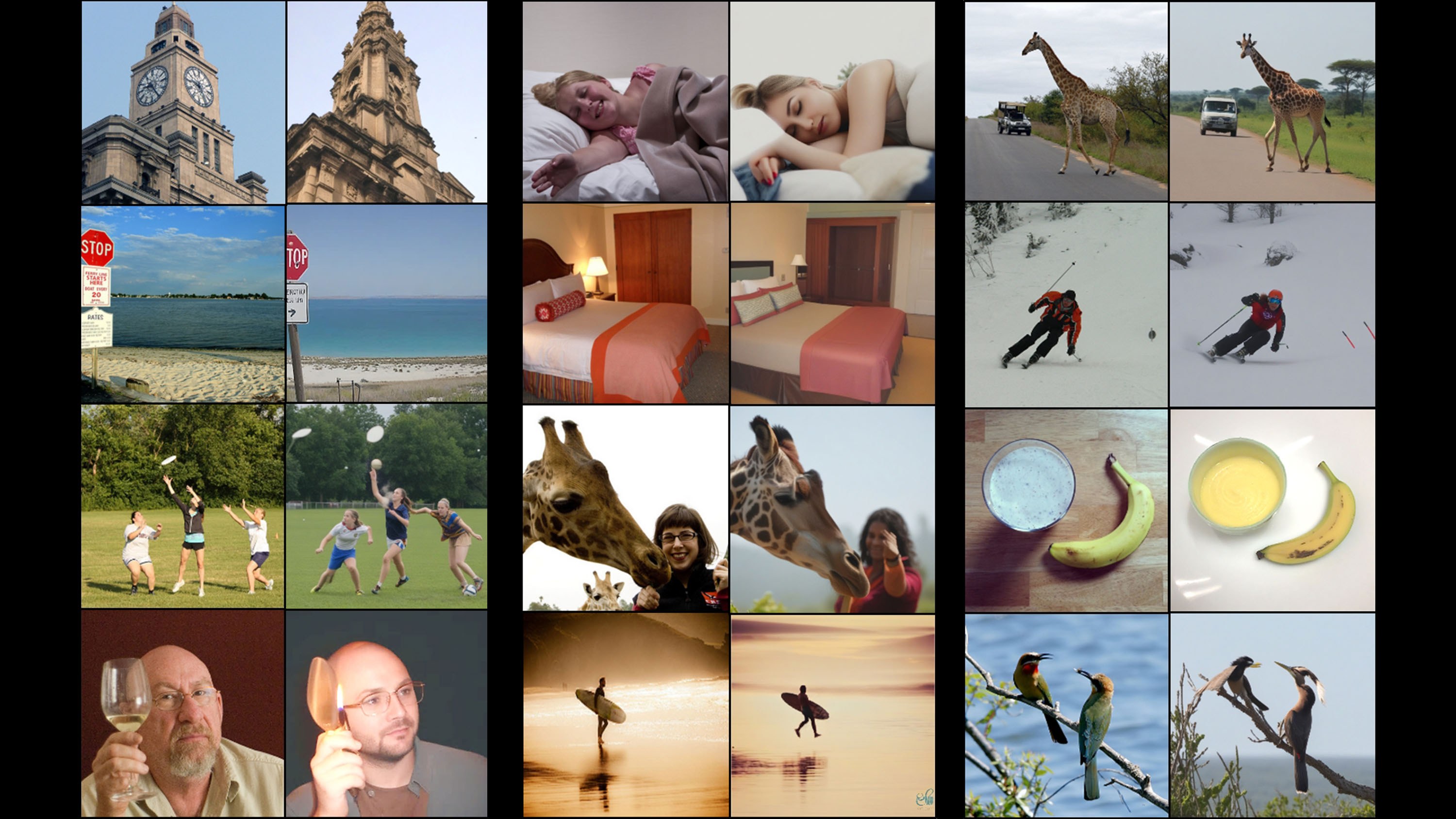
Neuroscientists have spent years attempting to decode functional magnetic resonance imaging data to visualize what a person sees, though early efforts typically yielded blurry, ambiguous shapes that looked more like Rorschach tests than actual objects. Recent progress in scanning resolution and machine learning architectures has finally shifted that baseline.
Michal Irani and her colleagues at the Weizmann Institute of Science in Rehovot, Israel, have developed an AI tool that decodes visual stimuli directly from human brain activity and reconstructs the viewed images with high fidelity. The system operates bidirectionally, allowing researchers both to reconstruct an image from brain signals and to simulate cortical responses from visual inputs.
Dual-Branch Decoding and Synthetic Training
The initial training phase relied on high-resolution fMRI datasets gathered from volunteers. Standard fMRI scanners capture active voxels covering around three cubic millimeters, with each cubic millimeter containing roughly 16,000 neurons. Irani and her team utilized higher-resolution equipment where each recorded voxel corresponded to approximately one cubic millimeter of neurons. To overcome the scarcity of paired neuroimaging data, the team trained an encoder alongside a two-branch decoder.
"Crucially, their 'brain decoder' has two branches—one to predict the structure of an image (where the colors are, for instance) and a second to predict its content"
One branch predicts the structural geometry and color distribution of the visual field, while the second resolves high-level semantic content, such as identifying specific objects on a plate. The encoder simulates fMRI responses for unindexed visual inputs, creating a synthetic training loop that enables the decoder to iteratively refine its output without collapsing into generic noise.
Calibration Efficiency and Ethical Implications
Practical application of brain-computer interfaces often encounters severe data-collection bottlenecks. Irani notes that her decoder requires calibration data from a new subject, though the method significantly lowers the acquisition barrier for downstream use cases compared to legacy models that demanded exhaustive retraining.
Medical applications represent an immediate operational horizon for such architectures. Judy Illes, a neuroethicist and professor of neurology at the University of British Columbia in Canada who was not involved in the research, describes the work as magnificent, noting that using this approach therapeutically to help individuals with neurologic conditions is tremendously exciting. Irani hopes the framework will assist locked-in patients with communication, provide mechanistic insights into cortical function, and potentially allow researchers to reconstruct the visual content of dreams.
However, advanced decoding precision introduces acute neuroethical concerns regarding mental privacy. Tommy Sprague, a neuroscientist at the University of California, Santa Barbara, warns that similar decoding frameworks could eventually be deployed to extract inner thoughts and mental imagery without an individual's explicit consent. While the method currently relies on high-resolution fMRI hardware, the algorithmic capability to resolve visual internal states highlights the urgent need to establish regulatory boundaries for neural data governance before non-invasive decoding frameworks mature further into commercial products.